A biosensor is an analytical device, used for the detection of a chemical substance, that combines a biological component with a physicochemical detector.[1][2][3][4] The sensitive biological element, e.g. tissue, microorganisms, organelles, cell receptors, enzymes, antibodies, nucleic acids, etc., is a biologically derived material or biomimetic component that interacts with, binds with, or recognizes the analyte under study. The biologically sensitive elements can also be created by biological engineering. The transducer or the detector element, which transforms one signal into another one, works in a physicochemical way: optical, piezoelectric, electrochemical, electrochemiluminescence etc., resulting from the interaction of the analyte with the biological element, to easily measure and quantify. The biosensor reader device connects with the associated electronics or signal processors that are primarily responsible for the display of the results in a user-friendly way.[5] This sometimes accounts for the most expensive part of the sensor device, however it is possible to generate a user friendly display that includes transducer and sensitive element (holographic sensor). The readers are usually custom-designed and manufactured to suit the different working principles of biosensors.
Biosensor system
A biosensor typically consists of a bio-receptor (enzyme/antibody/cell/nucleic acid/aptamer), transducer component (semi-conducting material/nanomaterial), and electronic system which includes a signal amplifier, processor & display.[6] Transducers and electronics can be combined, e.g., in CMOS-based microsensor systems.[7][8] The recognition component, often called a bioreceptor, uses biomolecules from organisms or receptors modeled after biological systems to interact with the analyte of interest. This interaction is measured by the biotransducer which outputs a measurable signal proportional to the presence of the target analyte in the sample. The general aim of the design of a biosensor is to enable quick, convenient testing at the point of concern or care where the sample was procured.[9][10][11]
Bioreceptors

In a biosensor, the bioreceptor is designed to interact with the specific analyte of interest to produce an effect measurable by the transducer. High selectivity for the analyte among a matrix of other chemical or biological components is a key requirement of the bioreceptor. While the type of biomolecule used can vary widely, biosensors can be classified according to common types of bioreceptor interactions involving: antibody/antigen,[12] enzymes/ligands, nucleic acids/DNA, cellular structures/cells, or biomimetic materials.[13][14]
Antibody/antigen interactions
An immunosensor utilizes the very specific binding affinity of antibodies for a specific compound or antigen. The specific nature of the antibody-antigen interaction is analogous to a lock and key fit in that the antigen will only bind to the antibody if it has the correct conformation. Binding events result in a physicochemical change that in combination with a tracer, such as fluorescent molecules, enzymes, or radioisotopes, can generate a signal. There are limitations with using antibodies in sensors: 1. The antibody binding capacity is strongly dependent on assay conditions (e.g. pH and temperature), and 2. the antibody-antigen interaction is generally robust, however, binding can be disrupted by chaotropic reagents, organic solvents, or even ultrasonic radiation.[15][16]
Antibody-antigen interactions can also be used for serological testing, or the detection of circulating antibodies in response to a specific disease. Importantly, serology tests have become an important part of the global response to the COVID-19 pandemic.[17]
Artificial binding proteins
The use of antibodies as the bio-recognition component of biosensors has several drawbacks. They have high molecular weights and limited stability, contain essential disulfide bonds and are expensive to produce. In one approach to overcome these limitations, recombinant binding fragments (Fab, Fv or scFv) or domains (VH, VHH) of antibodies have been engineered.[18] In another approach, small protein scaffolds with favorable biophysical properties have been engineered to generate artificial families of Antigen Binding Proteins (AgBP), capable of specific binding to different target proteins while retaining the favorable properties of the parent molecule. The elements of the family that specifically bind to a given target antigen, are often selected in vitro by display techniques: phage display, ribosome display, yeast display or mRNA display. The artificial binding proteins are much smaller than antibodies (usually less than 100 amino-acid residues), have a strong stability, lack disulfide bonds and can be expressed in high yield in reducing cellular environments like the bacterial cytoplasm, contrary to antibodies and their derivatives.[19][20] They are thus especially suitable to create biosensors.[21][22]
Enzymatic interactions
The specific binding capabilities and catalytic activity of enzymes make them popular bioreceptors. Analyte recognition is enabled through several possible mechanisms: 1) the enzyme converting the analyte into a product that is sensor-detectable, 2) detecting enzyme inhibition or activation by the analyte, or 3) monitoring modification of enzyme properties resulting from interaction with the analyte.[16] The main reasons for the common use of enzymes in biosensors are: 1) ability to catalyze a large number of reactions; 2) potential to detect a group of analytes (substrates, products, inhibitors, and modulators of the catalytic activity); and 3) suitability with several different transduction methods for detecting the analyte. Notably, since enzymes are not consumed in reactions, the biosensor can easily be used continuously. The catalytic activity of enzymes also allows lower limits of detection compared to common binding techniques. However, the sensor's lifetime is limited by the stability of the enzyme.
Affinity binding receptors
Antibodies have a high binding constant in excess of 10^8 L/mol, which stands for a nearly irreversible association once the antigen-antibody couple has formed. For certain analyte molecules like glucose affinity binding proteins exist that bind their ligand with a high specificity like an antibody, but with a much smaller binding constant on the order of 10^2 to 10^4 L/mol. The association between analyte and receptor then is of reversible nature and next to the couple between both also their free molecules occur in a measurable concentration. In case of glucose, for instance, concanavalin A may function as affinity receptor exhibiting a binding constant of 4x10^2 L/mol.[23] The use of affinity binding receptors for purposes of biosensing has been proposed by Schultz and Sims in 1979 [24] and was subsequently configured into a fluorescent assay for measuring glucose in the relevant physiological range between 4.4 and 6.1 mmol/L.[25] The sensor principle has the advantage that it does not consume the analyte in a chemical reaction as occurs in enzymatic assays.
Nucleic acid interactions
Biosensors employing nucleic acid based receptors can be either based on complementary base pairing interactions referred to as genosensors or specific nucleic acid based antibody mimics (aptamers) as aptasensors.[26] In the former, the recognition process is based on the principle of complementary base pairing, adenine:thymine and cytosine:guanine in DNA. If the target nucleic acid sequence is known, complementary sequences can be synthesized, labeled, and then immobilized on the sensor. The hybridization event can be optically detected and presence of target DNA/RNA ascertained. In the latter, aptamers generated against the target recognise it via interplay of specific non-covalent interactions and induced fitting. These aptamers can be labelled with a fluorophore/metal nanoparticles easily for optical detection or may be employed for label-free electrochemical or cantilever based detection platforms for a wide range of target molecules or complex targets like cells and viruses.[27][28] Additionally, aptamers can be combined with nucleic acid enzymes, such as RNA-cleaving DNAzymes, providing both target recognition and signal generation in a single molecule, which shows potential applications in the development of multiplex biosensors.[29]
Epigenetics
It has been proposed that properly optimized integrated optical resonators can be exploited for detecting epigenetic modifications (e.g. DNA methylation, histone post-translational modifications) in body fluids from patients affected by cancer or other diseases.[30] Photonic biosensors with ultra-sensitivity are nowadays being developed at a research level to easily detect cancerous cells within the patient's urine.[31] Different research projects aim to develop new portable devices that use cheap, environmentally friendly, disposable cartridges that require only simple handling with no need of further processing, washing, or manipulation by expert technicians.[32]
Organelles
Organelles form separate compartments inside cells and usually perform functions independently. Different kinds of organelles have various metabolic pathways and contain enzymes to fulfill its function. Commonly used organelles include lysosome, chloroplast and mitochondria. The spatial-temporal distribution pattern of calcium is closely related to ubiquitous signaling pathway. Mitochondria actively participate in the metabolism of calcium ions to control the function and also modulate the calcium related signaling pathways. Experiments have proved that mitochondria have the ability to respond to high calcium concentrations generated in their proximity by opening the calcium channels.[33] In this way, mitochondria can be used to detect the calcium concentration in medium and the detection is very sensitive due to high spatial resolution. Another application of mitochondria is used for detection of water pollution. Detergent compounds' toxicity will damage the cell and subcellular structure including mitochondria. The detergents will cause a swelling effect which could be measured by an absorbance change. Experiment data shows the change rate is proportional to the detergent concentration, providing a high standard for detection accuracy.[34]
Cells
Cells are often used in bioreceptors because they are sensitive to surrounding environment and they can respond to all kinds of stimulants. Cells tend to attach to the surface so they can be easily immobilized. Compared to organelles they remain active for longer period and the reproducibility makes them reusable. They are commonly used to detect global parameter like stress condition, toxicity and organic derivatives. They can also be used to monitor the treatment effect of drugs. One application is to use cells to determine herbicides which are main aquatic contaminant.[35] Microalgae are entrapped on a quartz microfiber and the chlorophyll fluorescence modified by herbicides is collected at the tip of an optical fiber bundle and transmitted to a fluorimeter. The algae are continuously cultured to get optimized measurement. Results show that detection limit of certain herbicide can reach sub-ppb concentration level. Some cells can also be used to monitor the microbial corrosion.[36] Pseudomonas sp. is isolated from corroded material surface and immobilized on acetylcellulose membrane. The respiration activity is determined by measuring oxygen consumption. There is linear relationship between the current generated and the concentration of sulfuric acid. The response time is related to the loading of cells and surrounding environments and can be controlled to no more than 5min.
Tissue
Tissues are used for biosensor for the abundance of enzymes existing. Advantages of tissues as biosensors include the following:[37]
- easier to immobilize compared to cells and organelles
- the higher activity and stability from maintaining enzymes in the natural environment
- the availability and low price
- the avoidance of tedious work of extraction, centrifuge, and purification of enzymes
- necessary cofactors for an enzyme to function exists
- the diversity providing a wide range of choices concerning different objectives.
There also exist some disadvantages of tissues, like the lack of specificity due to the interference of other enzymes and longer response time due to the transport barrier.
Microbial biosensors
Microbial biosensors exploit the response of bacteria to a given substance. For example, arsenic can be detected using the ars operon found in several bacterial taxon.[38]
Surface attachment of the biological elements

An important part of a biosensor is to attach the biological elements (small molecules/protein/cells) to the surface of the sensor (be it metal, polymer, or glass). The simplest way is to functionalize the surface in order to coat it with the biological elements. This can be done by polylysine, aminosilane, epoxysilane, or nitrocellulose in the case of silicon chips/silica glass. Subsequently, the bound biological agent may also be fixed—for example, by layer by layer deposition of alternatively charged polymer coatings.[39]
Alternatively, three-dimensional lattices (hydrogel/xerogel) can be used to chemically or physically entrap these (whereby chemically entrapped it is meant that the biological element is kept in place by a strong bond, while physically they are kept in place being unable to pass through the pores of the gel matrix). The most commonly used hydrogel is sol-gel, glassy silica generated by polymerization of silicate monomers (added as tetra alkyl orthosilicates, such as TMOS or TEOS) in the presence of the biological elements (along with other stabilizing polymers, such as PEG) in the case of physical entrapment.[40]
Another group of hydrogels, which set under conditions suitable for cells or protein, are acrylate hydrogel, which polymerizes upon radical initiation. One type of radical initiator is a peroxide radical, typically generated by combining a persulfate with TEMED (Polyacrylamide gel are also commonly used for protein electrophoresis),[41] alternatively light can be used in combination with a photoinitiator, such as DMPA (2,2-dimethoxy-2-phenylacetophenone).[42] Smart materials that mimic the biological components of a sensor can also be classified as biosensors using only the active or catalytic site or analogous configurations of a biomolecule.[43]
Biotransducer
Biosensors can be classified by their biotransducer type. The most common types of biotransducers used in biosensors are:
- electrochemical biosensors
- optical biosensors
- electronic biosensors
- piezoelectric biosensors
- gravimetric biosensors
- pyroelectric biosensors
- magnetic biosensors
Electrochemical
Electrochemical biosensors are normally based on enzymatic catalysis of a reaction that produces or consumes electrons (such enzymes are rightly called redox enzymes). The sensor substrate usually contains three electrodes; a reference electrode, a working electrode and a counter electrode. The target analyte is involved in the reaction that takes place on the active electrode surface, and the reaction may cause either electron transfer across the double layer (producing a current) or can contribute to the double layer potential (producing a voltage). We can either measure the current (rate of flow of electrons is now proportional to the analyte concentration) at a fixed potential or the potential can be measured at zero current (this gives a logarithmic response). Note that potential of the working or active electrode is space charge sensitive and this is often used. Further, the label-free and direct electrical detection of small peptides and proteins is possible by their intrinsic charges using biofunctionalized ion-sensitive field-effect transistors.[44]
Another example, the potentiometric biosensor, (potential produced at zero current) gives a logarithmic response with a high dynamic range. Such biosensors are often made by screen printing the electrode patterns on a plastic substrate, coated with a conducting polymer and then some protein (enzyme or antibody) is attached. They have only two electrodes and are extremely sensitive and robust. They enable the detection of analytes at levels previously only achievable by HPLC and LC/MS and without rigorous sample preparation. All biosensors usually involve minimal sample preparation as the biological sensing component is highly selective for the analyte concerned. The signal is produced by electrochemical and physical changes in the conducting polymer layer due to changes occurring at the surface of the sensor. Such changes can be attributed to ionic strength, pH, hydration and redox reactions, the latter due to the enzyme label turning over a substrate.[45] Field effect transistors, in which the gate region has been modified with an enzyme or antibody, can also detect very low concentrations of various analytes as the binding of the analyte to the gate region of the FET cause a change in the drain-source current.
Impedance spectroscopy based biosensor development has been gaining traction nowadays and many such devices / developments are found in the academia and industry. One such device, based on a 4-electrode electrochemical cell, using a nanoporous alumina membrane, has been shown to detect low concentrations of human alpha thrombin in presence of high background of serum albumin.[46] Also interdigitated electrodes have been used for impedance biosensors.[47]
Ion channel switch
The use of ion channels has been shown to offer highly sensitive detection of target biological molecules.[48] By embedding the ion channels in supported or tethered bilayer membranes (t-BLM) attached to a gold electrode, an electrical circuit is created. Capture molecules such as antibodies can be bound to the ion channel so that the binding of the target molecule controls the ion flow through the channel. This results in a measurable change in the electrical conduction which is proportional to the concentration of the target.
An ion channel switch (ICS) biosensor can be created using gramicidin, a dimeric peptide channel, in a tethered bilayer membrane.[49] One peptide of gramicidin, with attached antibody, is mobile and one is fixed. Breaking the dimer stops the ionic current through the membrane. The magnitude of the change in electrical signal is greatly increased by separating the membrane from the metal surface using a hydrophilic spacer.
Quantitative detection of an extensive class of target species, including proteins, bacteria, drug and toxins has been demonstrated using different membrane and capture configurations.[50][51] The European research project Greensense develops a biosensor to perform quantitative screening of drug-of-abuse such as THC, morphine, and cocaine [52] in saliva and urine.
Reagentless fluorescent biosensor
A reagentless biosensor can monitor a target analyte in a complex biological mixture without additional reagent. Therefore, it can function continuously if immobilized on a solid support. A fluorescent biosensor reacts to the interaction with its target analyte by a change of its fluorescence properties. A Reagentless Fluorescent biosensor (RF biosensor) can be obtained by integrating a biological receptor, which is directed against the target analyte, and a solvatochromic fluorophore, whose emission properties are sensitive to the nature of its local environment, in a single macromolecule. The fluorophore transduces the recognition event into a measurable optical signal. The use of extrinsic fluorophores, whose emission properties differ widely from those of the intrinsic fluorophores of proteins, tryptophan and tyrosine, enables one to immediately detect and quantify the analyte in complex biological mixtures. The integration of the fluorophore must be done in a site where it is sensitive to the binding of the analyte without perturbing the affinity of the receptor.
Antibodies and artificial families of Antigen Binding Proteins (AgBP) are well suited to provide the recognition module of RF biosensors since they can be directed against any antigen (see the paragraph on bioreceptors). A general approach to integrate a solvatochromic fluorophore in an AgBP when the atomic structure of the complex with its antigen is known, and thus transform it into a RF biosensor, has been described.[21] A residue of the AgBP is identified in the neighborhood of the antigen in their complex. This residue is changed into a cysteine by site-directed mutagenesis. The fluorophore is chemically coupled to the mutant cysteine. When the design is successful, the coupled fluorophore does not prevent the binding of the antigen, this binding shields the fluorophore from the solvent, and it can be detected by a change of fluorescence. This strategy is also valid for antibody fragments.[53][54]
However, in the absence of specific structural data, other strategies must be applied. Antibodies and artificial families of AgBPs are constituted by a set of hypervariable (or randomized) residue positions, located in a unique sub-region of the protein, and supported by a constant polypeptide scaffold. The residues that form the binding site for a given antigen, are selected among the hypervariable residues. It is possible to transform any AgBP of these families into a RF biosensor, specific of the target antigen, simply by coupling a solvatochromic fluorophore to one of the hypervariable residues that have little or no importance for the interaction with the antigen, after changing this residue into cysteine by mutagenesis. More specifically, the strategy consists in individually changing the residues of the hypervariable positions into cysteine at the genetic level, in chemically coupling a solvatochromic fluorophore with the mutant cysteine, and then in keeping the resulting conjugates that have the highest sensitivity (a parameter that involves both affinity and variation of fluorescence signal).[22] This approach is also valid for families of antibody fragments.[55]
A posteriori studies have shown that the best reagentless fluorescent biosensors are obtained when the fluorophore does not make non-covalent interactions with the surface of the bioreceptor, which would increase the background signal, and when it interacts with a binding pocket at the surface of the target antigen.[56] The RF biosensors that are obtained by the above methods, can function and detect target analytes inside living cells.[57]
Magnetic biosensors
Magnetic biosensors utilize paramagnetic or supra-paramagnetic particles, or crystals, to detect biological interactions. Examples could be coil-inductance, resistance, or other magnetic properties. It is common to use magnetic nano or microparticles. In the surface of such particles are the bioreceptors, that can be DNA (complementary to a sequence or aptamers) antibodies, or others. The binding of the bioreceptor will affect some of the magnetic particle properties that can be measured by AC susceptometry,[58] a Hall Effect sensor,[59] a giant magnetoresistance device,[60] or others.
Others
Piezoelectric sensors utilise crystals which undergo an elastic deformation when an electrical potential is applied to them. An alternating potential (A.C.) produces a standing wave in the crystal at a characteristic frequency. This frequency is highly dependent on the elastic properties of the crystal, such that if a crystal is coated with a biological recognition element the binding of a (large) target analyte to a receptor will produce a change in the resonance frequency, which gives a binding signal. In a mode that uses surface acoustic waves (SAW), the sensitivity is greatly increased. This is a specialised application of the quartz crystal microbalance as a biosensor
Electrochemiluminescence (ECL) is nowadays a leading technique in biosensors.[61][62][63] Since the excited species are produced with an electrochemical stimulus rather than with a light excitation source, ECL displays improved signal-to-noise ratio compared to photoluminescence, with minimized effects due to light scattering and luminescence background. In particular, coreactant ECL operating in buffered aqueous solution in the region of positive potentials (oxidative-reduction mechanism) definitively boosted ECL for immunoassay, as confirmed by many research applications and, even more, by the presence of important companies which developed commercial hardware for high throughput immunoassays analysis in a market worth billions of dollars each year.
Thermometric biosensors are rare.
Biosensor MOSFET (BioFET)
The MOSFET (metal-oxide-semiconductor field-effect transistor, or MOS transistor) was invented by Mohamed M. Atalla and Dawon Kahng in 1959, and demonstrated in 1960.[64] Two years later, Leland C. Clark and Champ Lyons invented the first biosensor in 1962.[65][66] Biosensor MOSFETs (BioFETs) were later developed, and they have since been widely used to measure physical, chemical, biological and environmental parameters.[67]
The first BioFET was the ion-sensitive field-effect transistor (ISFET), invented by Piet Bergveld for electrochemical and biological applications in 1970.[68][69] the adsorption FET (ADFET) was patented by P.F. Cox in 1974, and a hydrogen-sensitive MOSFET was demonstrated by I. Lundstrom, M.S. Shivaraman, C.S. Svenson and L. Lundkvist in 1975.[67] The ISFET is a special type of MOSFET with a gate at a certain distance,[67] and where the metal gate is replaced by an ion-sensitive membrane, electrolyte solution and reference electrode.[70] The ISFET is widely used in biomedical applications, such as the detection of DNA hybridization, biomarker detection from blood, antibody detection, glucose measurement, pH sensing, and genetic technology.[70]
By the mid-1980s, other BioFETs had been developed, including the gas sensor FET (GASFET), pressure sensor FET (PRESSFET), chemical field-effect transistor (ChemFET), reference ISFET (REFET), enzyme-modified FET (ENFET) and immunologically modified FET (IMFET).[67] By the early 2000s, BioFETs such as the DNA field-effect transistor (DNAFET), gene-modified FET (GenFET) and cell-potential BioFET (CPFET) had been developed.[70]
Placement of biosensors
The appropriate placement of biosensors depends on their field of application, which may roughly be divided into biotechnology, agriculture, food technology and biomedicine.
In biotechnology, analysis of the chemical composition of cultivation broth can be conducted in-line, on-line, at-line and off-line. As outlined by the US Food and Drug Administration (FDA) the sample is not removed from the process stream for in-line sensors, while it is diverted from the manufacturing process for on-line measurements. For at-line sensors the sample may be removed and analyzed in close proximity to the process stream.[71] An example of the latter is the monitoring of lactose in a dairy processing plant.[72] Off-line biosensors compare to bioanalytical techniques that are not operating in the field, but in the laboratory. These techniques are mainly used in agriculture, food technology and biomedicine.
In medical applications biosensors are generally categorized as in vitro and in vivo systems. An in vitro, biosensor measurement takes place in a test tube, a culture dish, a microtiter plate or elsewhere outside a living organism. The sensor uses a bioreceptor and transducer as outlined above. An example of an in vitro biosensor is an enzyme-conductimetric biosensor for blood glucose monitoring. There is a challenge to create a biosensor that operates by the principle of point-of-care testing, i.e. at the location where the test is needed.[73][74] Development of wearable biosensors is among such studies.[75] The elimination of lab testing can save time and money. An application of a POCT biosensor can be for the testing of HIV in areas where it is difficult for patients to be tested. A biosensor can be sent directly to the location and a quick and easy test can be used.
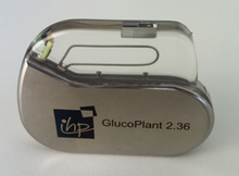
An in vivo biosensor is an implantable device that operates inside the body. Of course, biosensor implants have to fulfill the strict regulations on sterilization in order to avoid an initial inflammatory response after implantation. The second concern relates to the long-term biocompatibility, i.e. the unharmful interaction with the body environment during the intended period of use.[77] Another issue that arises is failure. If there is failure, the device must be removed and replaced, causing additional surgery. An example for application of an in vivo biosensor would be the insulin monitoring within the body, which is not available yet.
Most advanced biosensor implants have been developed for the continuous monitoring of glucose.[78][79] The figure displays a device, for which a Ti casing and a battery as established for cardiovascular implants like pacemakers and defibrillators is used.[76] Its size is determined by the battery as required for a lifetime of one year. Measured glucose data will be transmitted wirelessly out of the body within the MICS 402-405 MHz band as approved for medical implants.
Biosensors can also be integrated into mobile phone systems, making them user-friendly and accessible to a large number of users.[80]